JavaScript seems to be disabled in your browser. For the best experience on our site, be sure to turn on Javascript in your browser.
Tel: +1-832-696-8203
Email: [email protected]
Worldwide Distributors
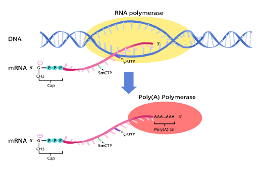
In vitro transcription of capped mRNA with modified nucleotides and Poly(A) tail
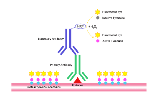
TSA (Tyramide Signal Amplification), used for signal amplification of ISH, IHC and IC etc.
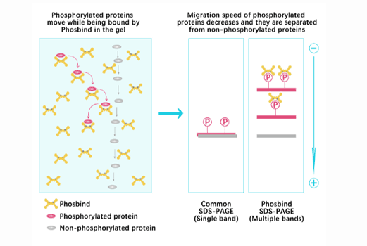
Separation of phosphorylated and non-phosphorylated proteins without phospho-specific antibody
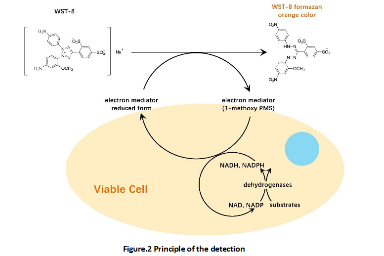
A convenient and sensitive way for cell proliferation assay and cytotoxicity assay
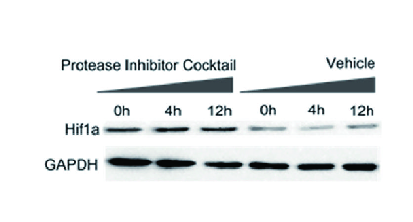
Protect the integrity of proteins from multiple proteases and phosphatases for different applications.
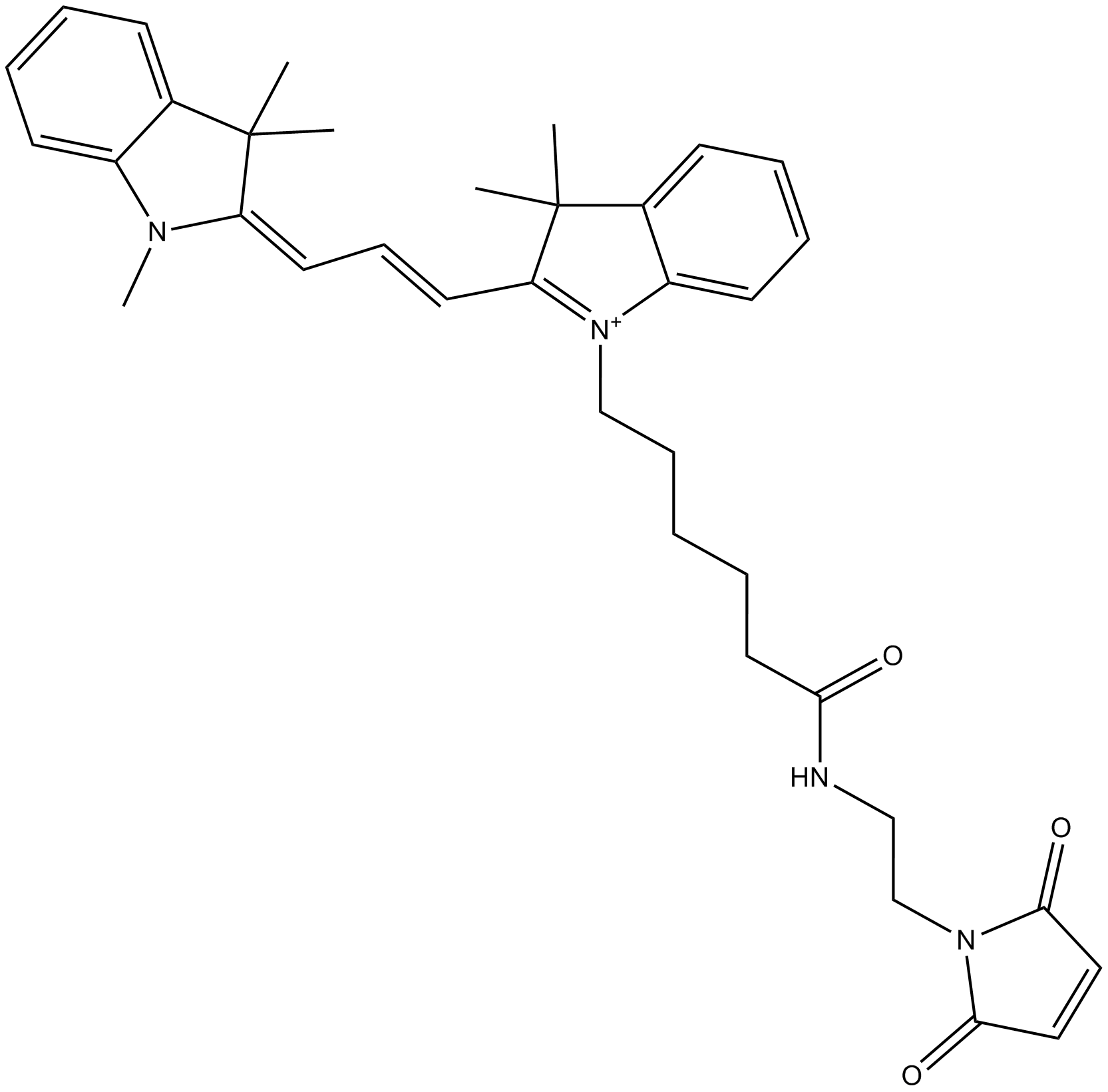
Cy3 maleimide is a selective and efficient fluorophore dye with low aqueous solubility. This labeling reagent can be used to attach Cy3 fluorophore to proteins and peptides which contain cysteine residues, as well as to other thiolate molecules (such as thiol-containing oligonucleotides). Before the labeling reactions, Cystines should be reduced with TCEP (tris-carboxyethylphosphine). For biomolecule labeling, the labeling reagent has low aqueous solubility, using of organic co-solvent to dissolve this molecular is necessary for efficient reaction. First, Cyanine dye should be dissolved in organic solvent and then added to a solution of biomolecule in appropriate aqueous buffer.
MSNs were labeled by Cy3 maleimide named MSN-Cy3s at the concentration of 10 μg ml-1. MSN-Cy3s incubated with MEF-mEGFP cells on a glass coverslip in culture medium for 2 h To further visualize the MSN locations in the cytosol we used super-resolution microscopy (3D SIM). The visually microscope image results confirm that MSNs were internalized by MEF-mEGFP cells [1].
Reference:[1]. Chiu, H.-Y.; Deng, W.; Engelke, H.; Helma, J.; Leonhardt, H.; Bein, T. Intracellular chromobody delivery by mesoporous silica nanoparticles for antigen targeting and visualization in real time. Scientific Reports, 2016, 6, 25019.


")