
ECL Chemiluminescent Substrate Detection Kit
- 1. Yeping Chen, Rongyuan Liang, et al. "Identification of ZNF652 as a Diagnostic and Therapeutic Target in Osteoarthritis Using Machine Learning." Journal of Inflammation Research Volume 17, 2025 - Issue
- 2. Ancong Xu, Fan Huang, et al. "Hyperbaric oxygen therapy attenuates heatstroke-induced hippocampal injury by inhibiting microglial pyroptosis." Int J Hyperthermia. 2024;41(1):2382162 PMID: 39043380
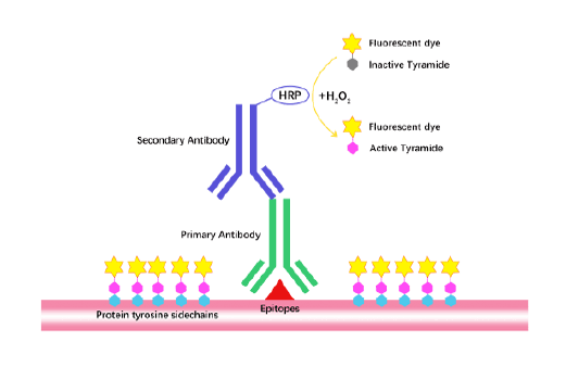
The core principle of ECL reagent detection is oxidation reaction luminescence: Under alkaline conditions, lumino, the main component of the luminescent substrate, is oxidized by H2O2 catalyzed by horseradish peroxidase (HRP) to generate 3-aminophthalene. The excited state intermediate of the acid emits photons when it returns to the ground state. The maximum emission wavelength is 425 nm. The photon signal can be captured by X-ray film or CCD imager.
ECL Chemiluminescent Substrate Detection Kit is used to detect antibodies that directly or indirectly label horseradish peroxidase (HRP) and its associated antigens. It is often used in WB detection and chemiluminescent immunodetection systems. The principle is that the protein or nucleic acid is transferred to the blotting membrane after electrophoresis, and the primary antibody and HRP-labeled secondary antibody bind to the target protein on the membrane, or the HRP-labeled probe directly or indirectly binds to the nucleic acid on the membrane. After washing the membrane, incubate the membrane with the ECL working solution prepared by this product for several minutes at room temperature, and fix the blotting membrane with plastic wrap and fix it on the X-ray exposure cassette. Then transfer to the dark room and press the X-ray film on the film to expose for several seconds to several hours. After development and fixation, the protein or nucleic acid bands can be clearly displayed on the X-ray film. It is also possible to directly scan the blot film without X-ray film exposure.

| Catalog No. | Name | Size |
| K1129 | ECL Chemiluminescent Substrate Detection Kit | 100 mL (50 mL each for A and B) |
Store at 2-8°C protected from light for two years. | ||