JavaScript seems to be disabled in your browser. For the best experience on our site, be sure to turn on Javascript in your browser.
Tel: +1-832-696-8203
Email: [email protected]
Worldwide Distributors
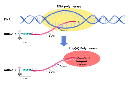
In vitro transcription of capped mRNA with modified nucleotides and Poly(A) tail
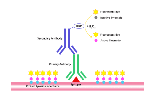
TSA (Tyramide Signal Amplification), used for signal amplification of ISH, IHC and IC etc.
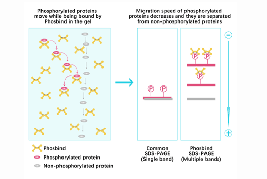
Separation of phosphorylated and non-phosphorylated proteins without phospho-specific antibody
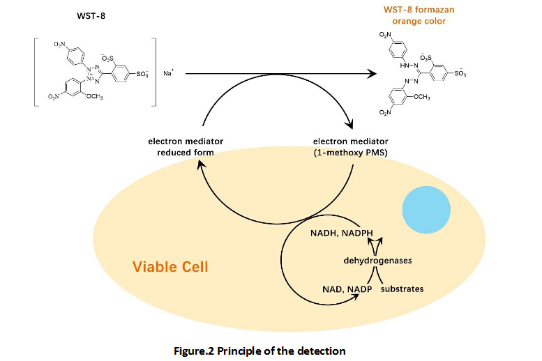
A convenient and sensitive way for cell proliferation assay and cytotoxicity assay
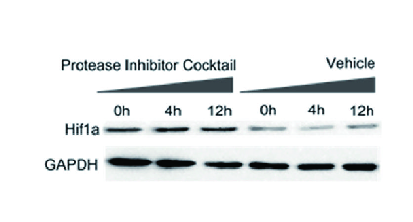
Protect the integrity of proteins from multiple proteases and phosphatases for different applications.
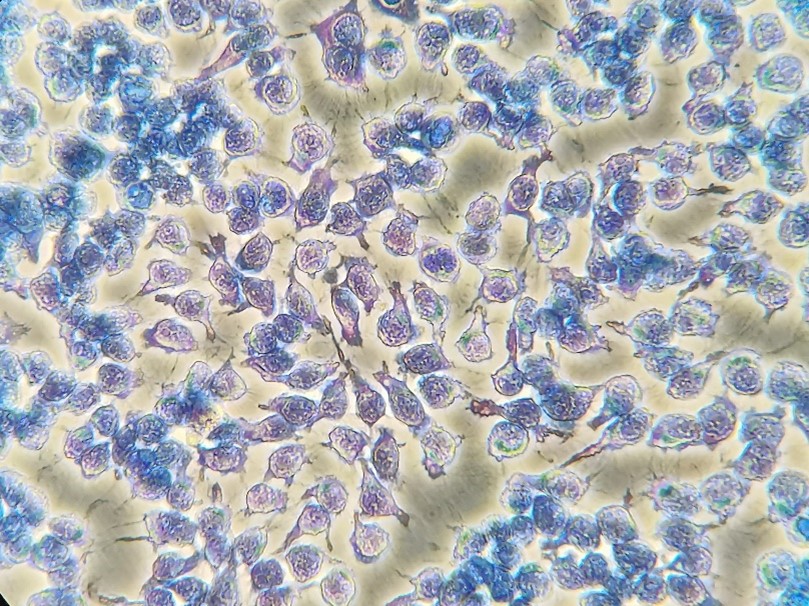
Giemsa Staining Solution is a mix of an anionic dye eosin, and a cationic dye azure. Giemsa staining is a widely used technique in histopathology and it acts as a golden standard for blood sample staining.
Eosin being acidic, bind to the mature red blood cells and eosinophils to give a pink color. While azure is basic and binds to acidic components like basophils, monocytes, and lymphocytes, producing a blue-purple or purple-red color. Neutrophils can react with eosin and azure together and are stained to pale purple. Each type of blood cell stains differently and can be distinguished morphologically. Similarly, this solution stains the basic cytoplasm pink, and stains the acidic nuclei blue-purple or purple-red. Giemsa staining is also used in karyotyping of the chromosomes. After Giemsa staining, chromosomes show different G banding and can be distinguished easily.
Giemsa staining solution is often used in combination with Wright staining solution.
The staining efficiency of this solution is shown in Figure 1.
Figure 1. Image of Hela cells stained with this solution
Store the components at room temperature away from light. Stable for at least 2 years.