
Protein A/G Magnetic Co-IP/IP Kit

Immunoprecipitation (IP) or Co-Immunoprecipitation (Co-IP). ) is a common technique for studying proteins or protein-protein interactions (PPIs) by using specific antibodies and mediators that bind to antibodies ( e.g., Protein A/G magnetic beads, etc.), or directly using a media conjugated to a specific antibody (such as agarose gel or magnetic beads) to isolate the protein of interest from the complex sample and can be used later SDS-PAGE or mass spectrometry analysis, etc.
This product is a classic magnetic bead method IP/Co-IP kit containing high-quality Protein A/G magnetic beads and optimized and validated immunoprecipitation necessary reagents, making IP or Co-IP experiments simpler, more convenient, and more convenient Highly efficient. Protein A/G can be specifically bound to the Fc terminal of the user's specific antibody and form Protein A/G magnetic beads after incubation for a certain period of time The antibody mixture (beads-Ab complex) is then added to the sample, which can be specifically recognized by the Fab terminal of the antibody to form Protein A/G magnetic beads - Antibody-antigen immune complex (Beads-Ab-Ag complex). Immune complexes are washed to remove unbound proteins, and bound immune complexes are then eluted from magnetic beads using methods such as acidic eluate or SDS-PAGE loading buffer for subsequent detection.
Protein A is a cell wall surface protein found in Staphylococcus aureus with a molecular weight of 42 kDa and is specifically associated with mammalian immunoglobulins (Immunoglobulin, Ig) Fc region will also bind to the Fab region of the human VH3 family. Protein G is a type C or G streptococcal bacteria The expressed immunoglobulin-binding protein specifically binds to the Mammalian immunoglobulin (Ig) FC region. This product is a modified recombinant Protein A (25 kDa) and Protein G (25 kDa), covalent with nanoscale amino magnetic beads Conjugation binding and retaining only the amino acid sequence associated with Fc terminal binding such as IgG removes sequences that may lead to non-specific binding outside the binding site, thereby effectively reducing non-specific binding. Protein A beads specifically bind to corresponding antibodies, such as human IgG1, IgG2, IgG4, mouse IgG2a, IgG2b and rabbit IgG, etc., and each Protein A molecule can bind 5 IgG molecules The antibodies that Protein G beads can bind to are human IgG1, IgG2, IgG3, IgG4, and mouse IgG1, IgG2a, IgG2b, IgG3, rat IgG1, IgG2a, IgG2b, IgG2c, as well as rabbit, goat polyclonal antibodies, etc., and each Protein G molecule can bind 3 IgG molecules. Protein A/G beads are mainly used for immunoprecipitation (IP) and co-precipitation (Co-IP). or Chromatin Immunoprecipitation (Ch-IP), and purification of antibodies in samples such as serum, cell culture supernatant, or ascites. The binding capacity of common immunoglobulin subclasses and the total binding capacity of different species are shown in the table below (Table 1).
Protein A/G beads is a 1:1 ratio configuration of Protein A beads and Protein G beads, which has a variety of significant advantages. First, high content and binding specificity of binding antibodies can be achieved. Compared with traditional Protein A/G agarose gels, this product has a smaller pore size, is less prone to non-specific adsorption, and has a high binding amount. 1 mL of magnetic bead suspension contains approximately 10 mg of magnetic beads and not less than 0.6 mg of recombinant Protein A/G, which can bind no less than 0.7 mg Human IgG, and the specific maximum binding amount is related to the type of antibody and the target protein. For experiments, efficient immunoprecipitation is typically performed using 10-20 μL of Protein A/G beads suspension for 500 μL samples. Second, ultra-fast binding of antibodies or antibody complexes can be achieved. Protein A/G beads (~200 nm) facilitate rapid and efficient binding of magnetic beads to antibodies or antibody complexes due to their large specific surface area. Usually, the adsorption process of antibodies or their complexes can be completed within 10 minutes, and the immunoprecipitation of the target protein can be completed within 30 minutes. Shortening the operation time can effectively avoid the degradation or denaturation of the target protein during long-term operation, and fully ensure the activity of the target protein. Due to the magnetic separation, IP and Co-IP can be performed each time compared to agarose gels by 40%. Finally, a variety of methods can be used to elute. Depending on factors such as the structure, biological function, and design requirements of the subsequent application of the protein of interest, a variety of eluents such as acidic solutions, SDS-PAGE loading buffers, or competitive peptides can be used for elution purposes. (See Table 2 for specific product parameters).
|
Species |
Subclass |
Protein A binding |
Protein G binding |
|
Human |
lgA |
++ |
- |
|
lgD |
++ |
- |
|
|
IgE |
++ |
- |
|
|
lgG1 |
++++ |
++++ |
|
|
lgG2 |
++++ |
++++ |
|
|
IgG3 |
- |
++++ |
|
|
lgG4 |
++++ |
++++ |
|
|
lgM |
++ |
- |
|
|
Mouse |
IgG1 |
+ |
++++ |
|
lgG2a |
++++ |
++++ |
|
|
lgG2b |
+++ |
+++ |
|
|
lgG3 |
++ |
+++ |
|
|
lgM |
+/- |
- |
|
|
Rat |
lgG1 |
- |
+ |
|
lgG2a |
- |
++++ |
|
|
lgG2b |
- |
++ |
|
|
lgG3 |
+ |
++ |
|
|
Avian egg yolk |
IgY |
- |
- |
|
Cow |
|
++ |
++++ |
|
Dog |
|
++ |
+ |
|
Goat |
|
- |
++ |
|
Guinea Pig |
lgG1 |
++++ |
++ |
|
lgG2 |
++++ |
++ |
|
|
Hamster |
|
+ |
++ |
|
Horse |
|
++ |
++++ |
|
Koala |
|
- |
+ |
|
Llama |
|
- |
+ |
|
Monkey (rhesus) |
|
++++ |
++++ |
|
Pig |
|
+++ |
+++ |
|
Rabbit |
|
++++ |
+++ |
|
Sheep |
|
+/- |
++ |
Table 1 affinity data for Protein A and Protein G for different sources and subtypes of IgG. ++++ = Strong Binding; ++~+++ = Medium Binding; + = Weak Binding; +/- = Weak or No Binding; - = No Binding.
|
Characteristics |
Description |
|
Product content |
10 mg/ mL magnetic beads in specific protective buffer |
|
Beads size |
~200 nm |
|
Magnetization |
Superparamagnetic |
|
Coupled protein |
Recombinant Protein A/G |
|
M.W. of protein |
~25 kDa (Protein A/G) |
|
Antibody concentration |
≥0.6 mg Protein A/G per mL beads |
|
Binding capacity |
≥ 0.7 mg human IgG per mL beads |
|
Specificity |
Antibodies from many different species, including mouse, human, rabbit, cow, goat, and sheep |
|
Elution method |
Elution with acid, competing peptide or SDS‐PAGE loading buffer |
|
Application |
IP, Co-IP, Protein purification |
Table 2 The main related indicators of Protein A/G beads.
| Components | K1309-10 T | K1309-50 T |
| Cell Lysis Buffer | 5 mL | 25 mL |
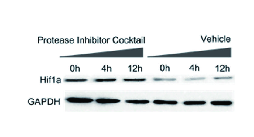
| Protease Inhibitor Cocktail (EDTA-Free,100X in DMSO) | 50 μL | 250 μL |
| 10X TBS | 5 mL | 30 mL |
| Neutralization Buffer | 100 μL | 500 μL |
| Acid Elution Buffer | 1 mL | 5 mL |
| Protein A/G beads | 200 μL | 1 mL |
| 5X Protein Loading Buffer (Reducing) | 200 μL | 1 mL |
Store Protease Inhibitor Cocktail (EDTA-Free,100X in DMSO) and 5X Protein Loading Buffer (Reducing) at -20°C and the rest of components at 4°C for 12 months. | ||