
Verteporfin
Verteporfin, derived from porphyrin, is a potent photosensitizing agent, which has all the features theoretically necessary for effective photodynamic therapy (PDT) in ocular neovascularization [1].
Experimentas showed that the mechanism of action of verteporfin therapy is intravascular damage leading to the formation of thrombus and selective vascular occlusion. Photodynamic therapy with verteporfin triggers cellular events consistent with a number of the changes described for cells rendered by different chemotherapeutic agents.
More than 90% of HL-60 cells treated with verteporfin at 100 ng/mL showed a hypodiploid level of DNA, when approximately 25% of irradiated cells treated with verteporfin at 50 ng/mL exhibited DNA fragmentation. For cells treated with verteporfin at 25 ng/mL, there was an 85% or greater loss in viability as determined by MTT assays performed 24 hours after irradiation. A 5–6 hours of the plasma half-life of verteporfin was shown in a pharmacokinetic studies in humans. At 6 mg/m2, which is the clinically relevant dose being used in neovascular AMD, no skin photosensitivity was detected at 24 hours, whereas at 18 mg/m2, baseline was reached by 5 days [1, 2].
Verteporfin was also found to inhibit autophagosome formation that does not require exposure to light. Verteporfin could target and modify p62 directly, a protein of scaffold and adaptor that binds both polyubiquitinated proteins destined for degradation and LC3 on autophagosomal membranes. Co-immunoprecipitation experiments demonstrated that crosslinked p62 oligomers retain their ability to bind to LC3 but show defective binding to polyubiquitinated proteins. Interestingly, small amounts of crosslinked p62 oligomers were detected in cells which were untreated, and other groups which were noted the accumulation of p62 forms with reduced SDS-PAGE mobility in cellular and animal models of oxidative stress and aging [3].
References:
[1].Schmidt-Erfurth U, Hasan T. Mechanisms of action of photodynamic therapy with verteporfin for the treatment of age-related macular degeneration. Survey Of Ophthalmology, 2000, 45(3): 195-214.
[2]. Granville DJ, Carthy CM, Jiang H, et al. Nuclear factor-kappa B activation by the photochemotherapeutic agent verteporfin. Blood, 2000, 95(1): 256-262.
[3]. Donohue E, Balgi AD, Komatsu M, et al. Induction of covalently crosslinked p62 oligomers with reduced binding to polyubiquitinated proteins by the autophagy inhibitor verteporfin. PLoS ONE, 2014, 9(12): e114964.
- 1. Peiqi Liu, Zhen Li, et al. "Mild heat stress promotes the differentiation of odontoblast-like MDPC-23 cells via yes-associated protein." Int J Hyperthermia. 2024;41(1):2369749. PMID: 38925872
- 2. Qingming Luo, et al. "Non-invasive modulation of meningeal lymphatics ameliorates ageing and Alzheimer's disease-associated pathology and cognition in mice." Nat Commun. 2024 Feb 16;15(1):1453. PMID: 38365740
- 3. Monica Y. Lee, et al. "Nucleoporin93 limits Yap activity to prevent endothelial cell senescence." Aging Cell. 2024 Feb 13:e14095. PMID: 38348753
- 4. Tung D Nguyen, Mihir K Rao, et al. "Nucleoporin93 (Nup93) Limits Yap Activity to Prevent Endothelial Cell Senescence." bioRxiv. 2023 Nov 14:2023.11.10.566598. PMID: 38014013
- 5. Li Ma, Qing Chang, et al. "Skull progenitor cell-driven meningeal lymphatic restoration improves neurocognitive functions in craniosynostosis." Cell Stem Cell. 2023 Nov 2;30(11):1472-1485.e7. PMID: 37863055
- 6. Vanessa Smer-Barreto, Andrea Quintanilla, et al. "Discovery of senolytics using machine learning." Nat Commun. 2023 Jun 10;14(1):3445. PMID: 37301862
- 7. Xiao Sun, Ping Sun, et al. "Melatonin alleviates doxorubicin-induced mitochondrial oxidative damage and ferroptosis in cardiomyocytes by regulating YAP expression." Toxicol Appl Pharmacol. 2022 Feb 15;437:115902. PMID: 35093381
- 8. Chen J, Wang L, et al. "Meningeal lymphatics clear erythrocytes that arise from subarachnoid hemorrhage." Nat Commun. 2020;11(1):3159. PMID:32572022
| Physical Appearance | A solid |
| Storage | Store at -20°C in the dark |
| M.Wt | 718.79 |
| Cas No. | 129497-78-5 |
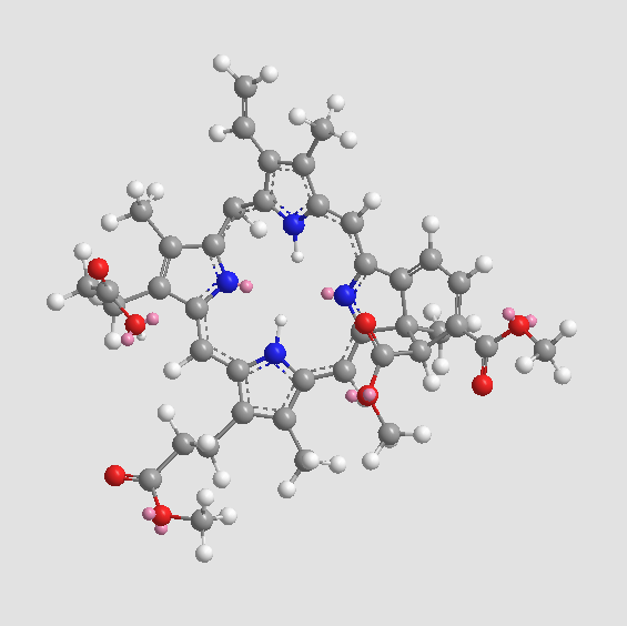
| Formula | C41H42N4O8 |
| Synonyms | CL 318952;Visudyne |
| Solubility | insoluble in EtOH; insoluble in H2O; ≥18.3 mg/mL in DMSO |
| SDF | Download SDF |
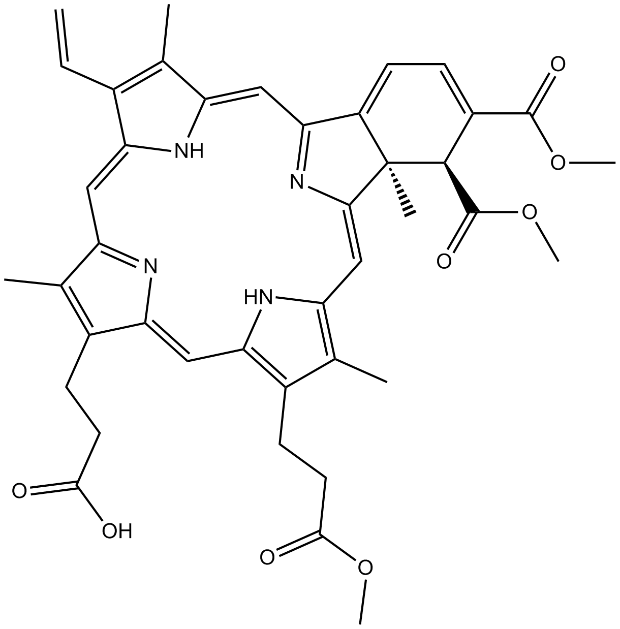
| Canonical SMILES | O=C([C@H]1[C@](/C(N=C2/C=C(C(C)=C/3C=C)\NC3=C/C4=N/C(C(CCC(O)=O)=C4C)=C\5)=C/C6=C(C)C(CCC(OC)=O)=C5N6)(C)C2=CC=C1C(OC)=O)OC |
| Shipping Condition | Small Molecules with Blue Ice, Modified Nucleotides with Dry Ice. |
| General tips | We do not recommend long-term storage for the solution, please use it up soon. |
| Cell experiment [1]: | |
|
Cell lines |
HL-60 cells |
|
Preparation method |
The solubility of this compound in DMSO is > 18.3 mg/mL. General tips for obtaining a higher concentration: Please warm the tube at 37 °C for 10 minutes and/or shake it in the ultrasonic bath for a while. Stock solution can be stored below - 20 °C for several months. |
|
Reacting condition |
0 ~ 100 ng/mL; 60 mins |
|
Applications |
Verteporfin at 50 ng/mL caused DNA fragmentation in 25% of irradiated cells. At lower concentrations of the photosensitizer, few (~ 5%) cells exhibited a hypodiploid amount of DNA. According to MTT assays performed 24 hrs after irradiation, there was an 85% or greater loss in viability for cells treated with Verteporfin at ≥ 25 ng/mL. Light or Verteporfin alone did not result in DNA fragmentation or affect cell viability. |
| Animal experiment [2]: | |
|
Animal models |
NOG mice bearing PhLO cells |
|
Dosage form |
140 mg/kg/day; s.c.; from days 22 to 28 |
|
Applications |
In NOG mice bearing PhLO cells, Verteporfin alone significantly reduced the leukemia cell ratio. The combination of Verteporfin and Dasatinib further reduced the number of leukemia cells in the spleen. In addition, there was no significant body weight loss in any group, which suggested that erteporfin and Dasatinib alone or in combination did not exhibited obvious drug toxicity. |
|
Other notes |
Please test the solubility of all compounds indoor, and the actual solubility may slightly differ with the theoretical value. This is caused by an experimental system error and it is normal. |
|
References: [1]. Granville DJ, Carthy CM, Jiang H, et al. Nuclear factor-kappa B activation by the photochemotherapeutic agent verteporfin. Blood, 2000, 95(1): 256-262. [2]. Schmidt-Erfurth U, Hasan T. Mechanisms of action of photodynamic therapy with verteporfin for the treatment of age-related macular degeneration. Survey Of Ophthalmology, 2000, 45(3): 195-214. |
|
| Description | Verteporfin is a potent second-generation photosensitizing agent derived from porphyrin. | |||||
| Targets | others | |||||
| IC50 | ||||||
Quality Control & MSDS
- View current batch:
Chemical structure
Related Biological Data

Related Biological Data

Related Biological Data

