JavaScript seems to be disabled in your browser. For the best experience on our site, be sure to turn on Javascript in your browser.
Tel: +1-832-696-8203
Email: [email protected]
Worldwide Distributors
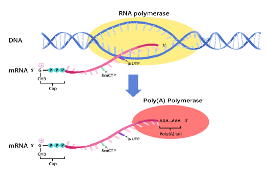
In vitro transcription of capped mRNA with modified nucleotides and Poly(A) tail
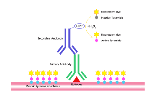
TSA (Tyramide Signal Amplification), used for signal amplification of ISH, IHC and IC etc.
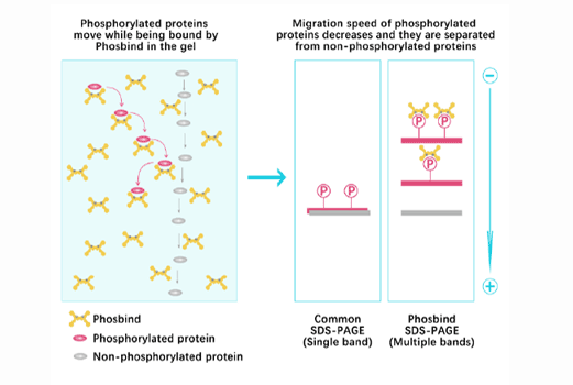
Separation of phosphorylated and non-phosphorylated proteins without phospho-specific antibody
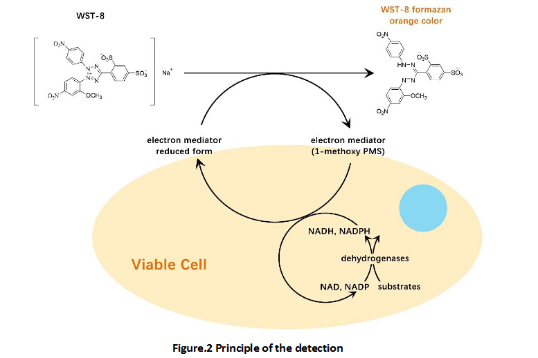
A convenient and sensitive way for cell proliferation assay and cytotoxicity assay
Protect the integrity of proteins from multiple proteases and phosphatases for different applications.
TGF-beta 1 (transforming growth factor beta 1) is one of three closely related mammalian members of the large TGF-beta superfamily that share a characteristic cystine knot structure. TGF-beta 1, -2 and -3 are highly pleiotropic cytokines that are proposed to act as cellular switches that regulate processes such as immune function, proliferation and epithelial-mesenchymal transition. Each TGF-beta isoform has some non‑redundant functions; for TGF-beta 1, mice with targeted deletion show defects in hematopoiesis and endothelial differentiation, and die of overwhelming inflammation. TGF‑ beta is activated from latency by pathways that include actions of the protease plasmin, matrix metalloproteases, thrombospondin 1 and a subset of integrins. Mature human TGF‑ beta 1 shares 100% aa identity with pig, dog and cow TGF‑ beta 1, and 99 % aa identity with mouse, rat and horse TGF‑beta 1.
Gene ID
7040
Accession #
P01137
Alternate Names
TGFB1, Human TGF-beta1, TGF-beta1, TGF beta1, TGFbeta1, h-TGF-beta1, rh-TGF-beta1, recombinant human TGF-beta1, recombinant TGF-beta, TGF.
Source
Chinese Hamster Ovary cell line, CHO
M.Wt
Apparent molecular mass of 24 kDa in SDS-PAGE under non-reducing conditions, 12 kDa under reducing conditions, a disulfide-linked homodimer of two 112 amino acid glycosylated polypeptide chains.
AA Sequence
Ala279-Ser390
Appearance
Sterile Filtered White lyophilized (freeze-dried) powder.
Stability & Storage
Use a manual defrost freezer and avoid repeated freeze-thaw cycles.
- 12 months from date of receipt, -20 to -70 °C as supplied.
- 1 month, 2 to 8 °C under sterile conditions after reconstitution.
- 3 months, -20 to -70 °C under sterile conditions after reconstitution.
Formulation
Lyophilized from 0.2 µm filtered concentrated solution in 35 % Acetonitrile and 0.1 % TFA.
Reconstitution
We recommend that this vial be briefly centrifuged prior to opening to bring the contents to the bottom. Reconstitute in sterile 4 mM HCl to a concentration of 0.1 mg/ml. Stock solutions should be apportioned into working aliquots and stored at ≤ -20 °C. Further dilutions should be made in appropriately buffered solutions.
Biological Activity
Measured by its ability to inhibit the IL-4-dependent proliferation of HT‑2 mouse T cells. The ED50 for this effect is 0.04-0.2 ng/mL. The specific activity of rHuTGF-β1 is approximately 2.5 × 10⁴ U/μg, which is calibrated against human TGF-β1 Standard.
Shipping Condition
Gel pack.
Handling
Centrifuge the vial prior to opening.
Usage
For Research Use Only! Not to be used in humans.